Rachel Doherty wint de LION Image Award met viraal microbootje
De jaarlijkse LION Image award gaat naar de foto van een 30 micrometer lang 3D-geprint bootje dat in oktober viraal ging, ingediend door Rachel Doherty van de groep van Daniela Kraft.
De jaarlijkse beeldwedstrijd voor Leidse natuurkundigen trok in dit corona-jaar slechts zes inzendingen, maar de kwaliteit en de diversiteit in onderwerpen was wel hoog (zie onder voor de beelden met de Engelstalige toelichting van de inzenders).
De winnaar werd volgens een beproefd recept bepaald door de secretaresses van de vakgroepen van het LION, die stemden voor de zeer herkenbare 'kleinste boot ter wereld', een 30 micrometer lang, 3d-geprint speelgoedbootje.
Na de online aankondiging gaf winnares Rachel Doherty een kort praatje over het beeld en de kleine mediahausse die het in oktober 2020 veroorzaakte. Het elektronenmicroscopiebeeld haalde talloze internationale media, waaronder de BBC, de Spaanse televisie, NRC Handelsblad, Engadged en zelfs twee Arabische scheepvaartnieuwswebsites.
'Hij heet '3D Benchy', en het is een ontwerp dat gebruikt wordt om 3D-printers te testen', legde Doherty uit. Zij is de labmanager en technicus die hielp het beeld te maken en die het ook inzond. De 3D-printer was de nieuwe Nanoscribe Photonic Professional, die in staat is om ontwerpen op een schaal van micrometers uit te printen. 'Dit is niet eens de kleinste boot die we geprint hebben', onthulde Doherty, 'we hebben er ook een paar van 10 micrometer gemaakt.' 10 micrometer is ongeveer een tiende van de dikte van een haar.
Bootje vaart ook echt
Ook liet Doherty een video zien waarin het bootje ook echt vaart. Een deel van het bootje is bedekt met een dunne laag platina, waardoor het bootje 'vaart' in een waterstofperoxide-oplossing, voortgestuwd door een chemische reactie van het platina.
De onderzoeksgroep van Doherty en Kraft doet onderzoek naar microzwemmers, ofwel kleine deeltjes die bewegen in vloeistoffen als water, en die je kunt volgen met hulp van een microscoop.
Doelen zijn onder andere beter begrip van biologische microzwemmers, zoals bacteriën, of het ontwerpen van machines en robots op micrometerschaal. Tegelijkertijd met de bootjes werden vele andere vomen geprint, zoals schroef-achtige spiralen.
De winnaar van de LION Image Award ontvangt 100 euro, roem bij vakgenoten, en het beeld zal vertoond worden op de wanden van het instituut, die de laatste tijd helaas weinig bekijks hebben.

THE WINNER: Rachel Doherty - The world's smallest boat
This microboat is 30 micrometres long, about a third of the thickness of a human hair, and can propel itself in solution using a chemical reaction. It was 3D printed and coated with a layer of platinum. The boat uses hydrogen peroxide as fuel, which is broken down by the platinum coating pushing the boat forward. The image was taken with an electron microscope. Submitted by Rachel Doherty
The Runners-Up

Ali Azadbakht - Trapped Particles
Optical tweezers are real versions of the tractor beam in Star Trek. They employ highly-focused laser beams to trap micrometer sized particles. Arthur Ashkin won the 2018 Nobel Prize for the invention of optical tweezers and their application in biology.
By diffracting the laser beam, we produced many traps simultaneously, and individually controlled them with nanometer resolution. Here, twenty-eight particles are trapped to form the letters LION (Leids Instituut voor Onderzoek in de Natuurkunde). Submitted by Ali Azadbakht

Gal Lemut - Majorana Fermions
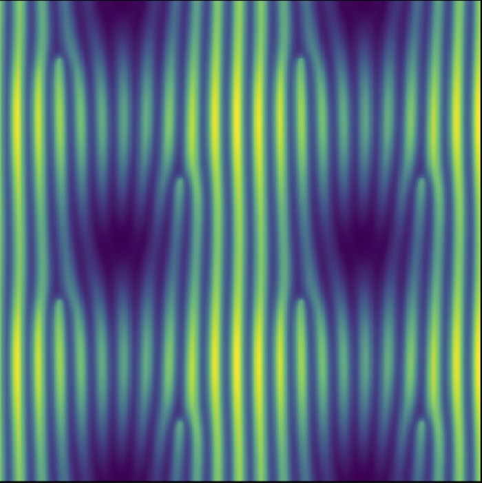
Majorana fermions are a special type of quasiparticles which are their own antiparticles. They are formed as a superposition of electrons and holes. This figure shows Majorana fermions formed in a superconductor under the action of a magnetic field.
In such systems, they can form special extended Landau level states. The oscillating pattern in their wave function amplitude reveals the interference of the electron and hole components. Submitted by Gal Lemut

Peter Neu - Meandering Carbon Nanotubes
Each carbon nanotube is only 2 nanometers in diameter and about 10 μm long, with a wall consisting of a single atomic layer of graphene. The carbon nanotubes were dispersed in water at a high concentration. When deposited on silicon and left to dry, they bundle together and form meandering abstract shapes.
Towards the top of the picture, we see bundles splitting up, which happens after prolonged exposure to the electron beam of the Scanning Electron Microscope (SEM). The field of view is 140 μm wide. Submitted by Peter Neu

Gallium Bombarded Rice Paddies - Remko Fermin
This false coloured electron microscope image might remind you of terraced rice fields that lie above steep cliffs. The unconventional scale bar, measured in lengths of the Covid-19 virus, reveals that we are dealing with a totally different scale. This image was taken after focused ion-beam processing, during which a sample is bombarded with highly energetic Gallium ions, to sculpt it into a microstructure, similar to a jigsaw. 'We needed very high ion currents on this specific sample since the crystal was relatively large, resulting in these randomly formed terraces at the edges (green) and pillars on the side (orange) of the crystal', says Remko Fermin who submitted the picture.

Tobias A. de Jong - Atomic Moiré Phases
When combining two atomic lattices with a slight twist, a moiré pattern shows up. This moiré pattern will, quite literally, magnify any shifts between the two atomic lattices. By comparing a Low Energy Electron Microscopy (LEEM) image of the moiré pattern with a reference wave, we can extract the local phase and obtain the local relative shifts of the atomic lattices with a very high precision. Here, this relative phase is shown on top of the moiré pattern of a graphene layer on top of an hexagonal boron nitride layer, curving around a fold in the graphene. Submitted by Tobias A. de Jong
